Visit profile

COMBS Project

COMBS
The cochlea is part of the auditory system and, as such, it is a very important part of the inner ear. It is responsible for the transfer of audio signals to the brain. Specific disorders of the cochlea can be solved only with a special implant. This implant is an electronic device which simulates the functionality of the cochlea.
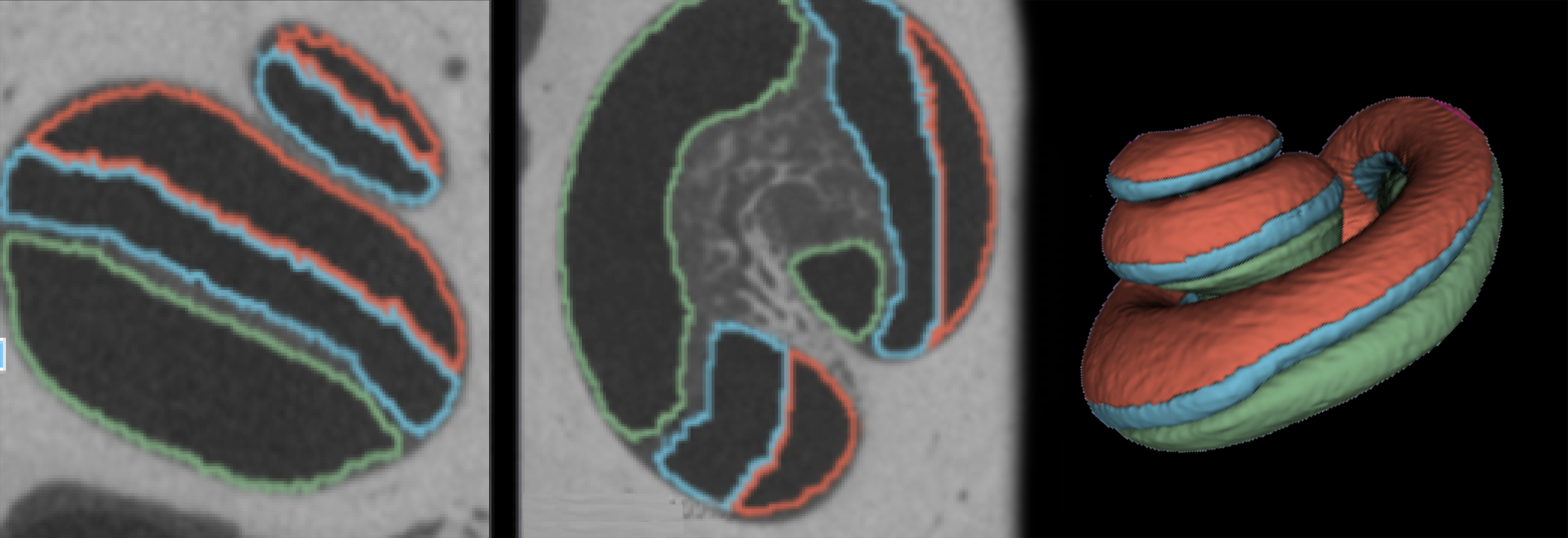
A successful cochlea surgery requires appropriate medical images to facilitate the doctors’ decision-making in finding the right type of implant and the suitable size of the cochlea implant. Due to the particularly delicate structure of the cochlea, a problem emerges: The required resolution for a metric description is namely inferior to the technical resolution of medical images such as CT and MRI. In order to improve image resolution, two main methods are used in this project: a model-based segmentation and the production of a very precise histological model. Thanks to this method, three goals can be achieved:
1. The segmentation of the cochlea, with a resolution that is far above CT / DVT resolution.
2. The recognition of the chambers within the cochlea (scalae).
3. The identification of metric parameters, including the distance (minimum to maximum).


